Measurement
Reading a shape-shifting protein directly in solution, fast enough to catch structure that lasts only milliseconds.
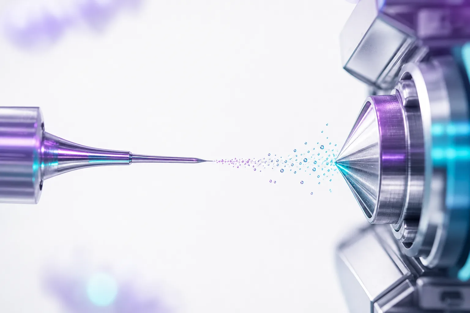
Reading a shape-shifting protein directly in solution, fast enough to catch structure that lasts only milliseconds.
Read a disordered protein where it lives, in solution and in motion, fast enough to catch the structure that appears for only a moment.
Hydrogen-deuterium exchange mass spectrometry gives Peptone a residue-level read on proteins that hold no fixed shape. Backbone amide hydrogens trade places with deuterium from the solvent, and the extra mass they carry records how protected each region is.
The states that matter most in a disordered protein are the ones that barely exist, transient folds that open a binding site for a few milliseconds before dissolving. Peptone's ultra-fast mixing reaches that window, measuring exchange below fifty milliseconds where conventional HDX-MS is still blind.
Every backbone amide carries a hydrogen that can swap for deuterium when the solvent reaches it. Amides that transiently fold or hydrogen-bond are shielded and exchange slowly, while exposed amides exchange fast. Mass spectrometry weighs the result, so the deuterium taken up over time becomes a residue-resolved map of protection across the whole chain, measured directly in solution with no crystal or fixed structure required.
Intrinsically disordered proteins exchange almost as fast as free peptides, so most of the informative signal is gone within the first moments of labelling. Peptone mixes protein and deuterated buffer in a continuous-flow microfluidic device with a dead time under a millisecond, then sets the labelling time by how far the stream travels before it is quenched. This puts the full sub-fifty-millisecond regime within reach, where conventional manual and robotic workflows only begin to sample after seconds.
A transient fold protects its amides only briefly before they exchange away. By the time a classical experiment takes its first measurement, that difference has washed out and the region looks fully disordered. Reading the millisecond window separates a transiently structured region from bulk disorder, exposing the low-population, binding-competent states that are invisible to slower methods yet central to how disordered targets are drugged.
Our D-TRON workflow turns these exchange rates into quantitative structural constraints that feed directly into ensemble modeling. Instead of a static picture, HDX-MS gives the platform experimental evidence about which conformations a target actually samples, and how a ligand shifts that balance.
A three-stage figure. First, a disordered backbone in deuterated buffer swaps amide hydrogens for deuterium and its mass spectrum shifts to higher mass. Next, a continuous-flow microfluidic mixer labels the protein in under a millisecond and quenches it across the sub-fifty-millisecond window. Finally, a deuterium-uptake curve shows a transiently protected region separating from bulk disorder only inside that early window, long before a conventional HDX measurement would begin.